What Are 3D Dental Scans?
3D dental scans, also known as cone beam computed tomography (CBCT), create detailed three-dimensional images of your teeth, soft tissues, nerve pathways, and bone in a single scan. Unlike traditional dental X-rays that produce flat, two-dimensional images, 3D scans capture hundreds of images from multiple angles and combine them into a comprehensive digital model.
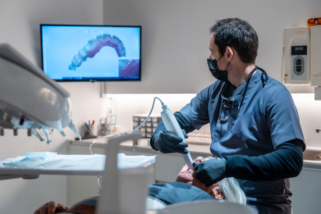
The scanner rotates around your head in a complete circle, taking images as it moves. Specialized software then reconstructs these images into a three-dimensional representation your dentist can examine from any angle. This process takes only seconds to complete yet produces incredibly detailed results.
How 3D Imaging Differs from Traditional X-Rays
Traditional X-rays compress three-dimensional structures into flat images, which can cause overlapping and make certain problems difficult to identify. A cavity hiding behind a tooth or a hairline crack in a root may not appear clearly on standard X-rays. With 3D dental scans, your dentist can rotate the image, zoom in on specific areas, and view cross-sections of any structure. This capability makes finding hidden issues much easier and helps create more accurate treatment plans.
“The chair has an extendable arm (C-arm) while the table has a rotator (gantry) that both rotate 360 degrees around the patient’s head, taking multiple images at once.”
Benefits of Trusted 3D Dental Scans in Sunnyvale
Choosing a dental practice that offers 3D imaging provides several advantages for your oral health care. The technology improves nearly every aspect of diagnosis and treatment planning.
Accuracy stands as the primary benefit of 3D dental scans. Measurements taken from these scans are precise to fractions of a millimeter, which becomes particularly important for procedures like dental implant placement. Knowing the exact width, depth, and density of your jawbone allows for implants to be positioned perfectly the first time.
- Treatment planning improves dramatically when dentists can see every angle of a problem before beginning work
- Hidden infections, cysts, or tumors become visible that might otherwise go undetected
- Root canal procedures become more predictable when the full anatomy of tooth roots is clearly visible
- Orthodontic treatment benefits from seeing exactly how teeth and roots are positioned relative to each other
- Surgical procedures carry less risk when the exact location of nerves and sinuses is known beforehand
The reduced radiation exposure compared to medical CT scans also makes 3D dental imaging a safer diagnostic option. Modern cone beam technology focuses the X-ray beam precisely on the area being examined, minimizing unnecessary exposure.
“The X-rays are compiled using a series of algorithms to furnish high-resolution 3D images.”
When You May Need a 3D Dental Scan
Your dentist may recommend a 3D scan for various situations where additional detail would improve your care. Planning for dental implants represents one of the most common uses, as the scan reveals whether you have sufficient bone density and shows exactly where nerves and sinuses are located. Patients considering Invisalign or other orthodontic treatments also benefit from the detailed view of tooth positions and root structures.
Additional Uses for 3D Imaging
Complex extractions, including wisdom teeth removal, become safer when the dentist can see the exact position of roots and nearby nerves. Diagnosing jaw joint disorders, evaluating the extent of periodontal disease, and locating the source of unexplained dental pain all benefit from 3D imaging capabilities. If you have experienced facial trauma, a 3D scan can reveal fractures or damage that traditional X-rays might miss.
“They can evaluate diseases of the jaw, dentition, body structures of the face, nasal cavity, and sinuses.”
What to Expect During Your 3D Scan
The scanning process is quick, comfortable, and completely painless. You will stand or sit while a small arm rotates around your head, capturing images. The entire process typically takes less than one minute. You do not need to enter an enclosed space as with medical CT scanners, which makes the experience comfortable even for those who feel anxious in small spaces.
No special preparation is required before your scan. You will simply remove any jewelry, glasses, or metal objects from your head and neck area. Our team will position you carefully to ensure the clearest possible images, and then you simply remain still for the brief scanning period.
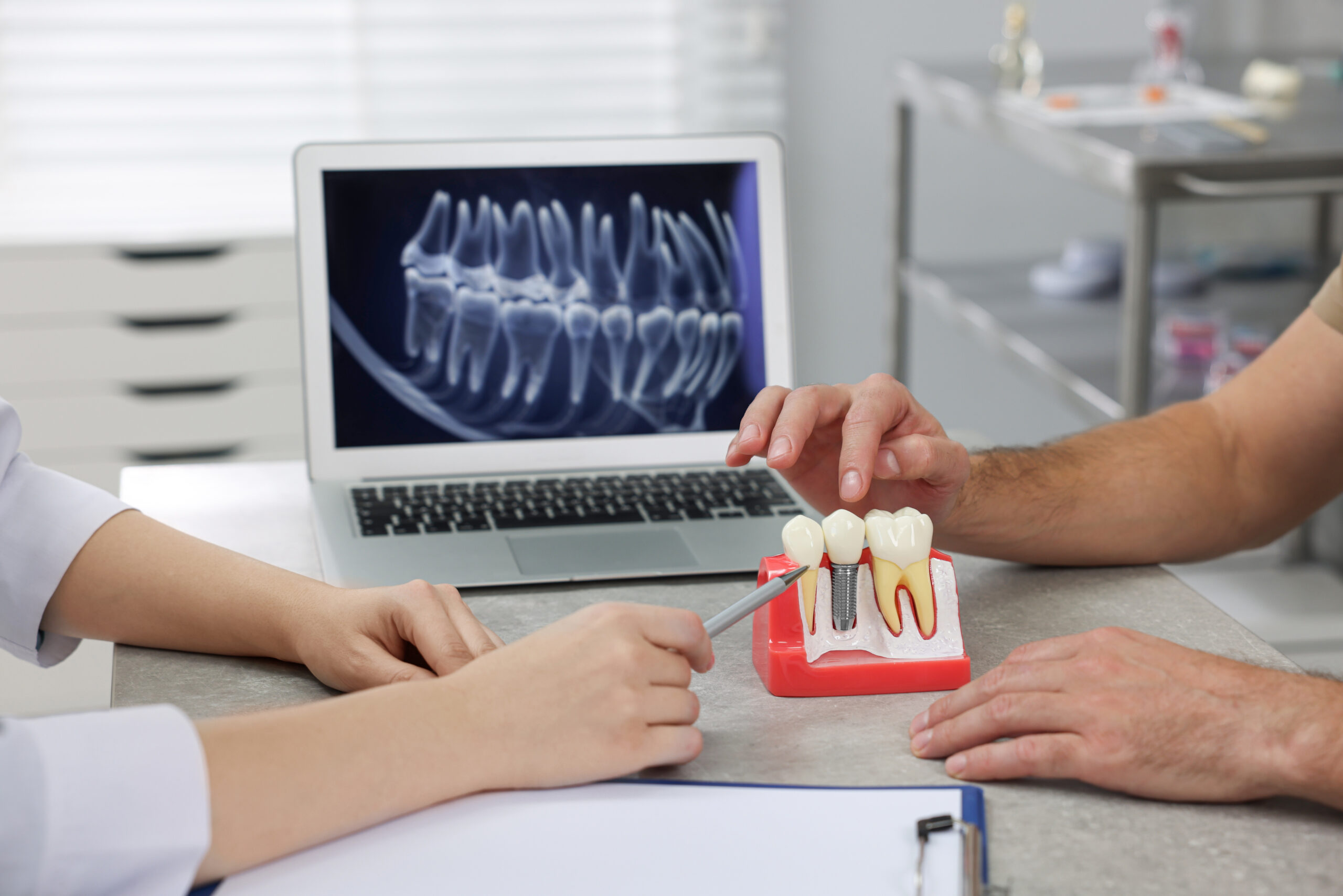
“The patient will be able to see their 3D images and follow the dentist as they move through the treatment plan, pointing at the treatment areas.”
Questions Answered on This Page
Q. How does a 3D cone beam work?
Q. What is the difference between traditional and cone beam scanners?
Q. What can a cone beam scan reveal about a patient?
People Also Ask
Q. What does the dentist look for in a dental examination?
Q. Who should get tooth fillings?
Q. How do lifestyle choices affect dental health?
Q. What should patients do if they have sensitive teeth?
Q. How can I find out if my employer’s plan covers dental treatments?
| Experience precise 3D dental imaging in Sunnyvale — request your appointment today. | Request an Appointment |
Frequently Asked Questions About 3D Cone Beam Scans
Q. Are cone beam scans covered by insurance?
A.Cone beam scans are increasingly being covered by insurance companies as there is a rise in the use of these scans with daily patients. However, not all insurance providers cover these types of scans. We recommend contacting your insurance provider to understand the details of your plan or request coverage for a cone beam scan.
Q. What kind of image does a cone beam scanner produce?
A.Cone beam scanners not only take images of the mouth but also the entire head. The positioning of your teeth affects the entire head, allowing dentists to see the underlying bone structure and jawline and creating more accurate treatments. They are especially useful for procedures around delicate areas and for planning correctional procedures.
Q. How long have cone beams been used?
A.Cone beam scanners are fairly new and not all dental clinics use them. Although they were invented in 1967, they were first introduced in the European market in 1996 and in the U.S. market in 2001. Today, about 72% of dentists use CBCT scanners.
Q. How safe are CBCT scans?
A.CBCT scans emit 200-300 times less or 1/10 the amount of radiation as traditional imaging systems and X-rays. Cone beam CT scanners also allow dentists to emit a certain dosage, making them safer and much more flexible. However, they still emit a certain amount of radiation and can be considered not safe, especially for children, pregnant women, and the elderly. Regardless, all X-ray scans in dentistry require some amount of radiation, and CBCT scans emit the lowest.
Q. Is there anything I should prepare for or bring for a CBCT exam?
A.There is no preparation required before a CBCT scan. It is important to remove all loose or metallic objects before the exam, as with any other X-ray exam. Patients with metal implants can get a CBCT scan. The dentist will ask you to stay still and do not swallow or talk during the exam.
Schedule Your Appointment with Smile Craft Dental
Finding the best 3D dental scans in Sunnyvale starts with choosing a dental practice committed to using current technology and providing patient-centered care. Smile Craft Dental combines advanced imaging capabilities with a team that takes time to explain findings and answer your questions. Our beautiful office and highly rated patient care make every visit comfortable, and Spanish-speaking staff members are available to assist patients who prefer communication in Spanish.
Ready to experience the difference advanced dental technology makes? Contact our team to schedule your appointment at our Sunnyvale location or learn more about how 3D dental scans may benefit your treatment. Our dedicated team looks forward to providing you with the comprehensive, thoughtful dental care you deserve.
 Scan here to view this page, 3D Cone Beam and 3D Dental Scans, on mobile
Dental Terminology
Dental Cone Beam Computed Tomography (CBCT)A dental cone beam computed tomography (CBCT) is a special type of X-ray equipment that reconstructs a 3D image of the patient’s anatomy.
DentistryDentistry is the profession that deals with preventing and treating any diseases and aberrations of the teeth, gums, and oral cavity.
Helpful Related Links
- American Dental Association (ADA). Glossary of Dental Clinical Terms. 2023
About our business, license, and website security
- Smile Craft Dental was established in 2019.
- We accept the following payment methods: American Express, Cash, Check, Discover, MasterCard, and Visa
- We serve patients from the following counties: San Mateo County and Santa Clara County
- We serve patients from the following cities: Redwood City, San Carlos, Atherton, Redwood Shores, Menlo Park, Woodside, Palo Alto, East Palo Alto, Belmont, and San Mateo
- Norton Safe Web. View Details
- Trend Micro Site Safety Center. View Details

Back to top of 3D Cone Beam and 3D Dental Scans